
Incubator Accessories

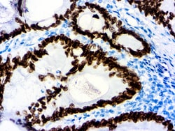
Diagnostic Biosystems SATB2 SATB2 (EP 281)
Special AT-rich sequence-binding protein 2 (SATB2) is a recently described marker that functions as a nuclear matrix-associated transcription factor. It has been reported that SATB2, in combination with CK20, could identify almost all colorectal carcinomas, including poorly differentiated colorectal carcinomas. Upper gastrointestinal (GI) carcinomas and pancreatic ductal carcinomas are usually negative for SATB2, and ovarian carcinomas, lung adenocarcinomas, and adenocarcinomas from other origin are rarely positive for SATB2. Therefore, SATB2 is a good marker for identifying a carcinoma of colorectal origin when working on a tumor of unknown primary. It can help differentiate colorectal metastasis (SATB2+) from primary pulmonary adenocarcinoma of mucinous or enteric type (SATB2- but often CDX2+)\n


Diagnostic Biosystems DAB GONE KIT Montage DAB Gone™ Kit
Montage DAB GoneTM removes diaminobenzidine (DAB) stains and precipitate from surfaces. Contamination with DAB residue can be a problem that may affect staining quality or automated immunohistochemistry stainers. Bleach will only reduce the color intensity of DAB stains, whereas,Montage DAB GoneTM removes the residual DAB stain that may clog instrument probes and tubing. This kit can also be used to clean glassware and other labware used in the lab that has been exposed to DAB and has retained DAB stains even after normal cleaning. It chemically reacts with DAB to inactivate the carcinogenic properties.\n\nThis kit has been designed to clean DAB from Diagnostic BioSystems MontageTM 360. This kit can also be used with instruments such as: BioCare NemesisTM, intelliPATHTM, Dako Autostainer, Autostainer Plus, Autostainer Plus Link, Lab Vision AutostainerTM 360, 480, and 720 models.
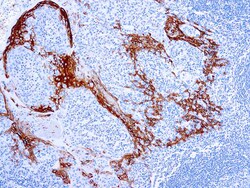
Diagnostic Biosystems CYTOKERATIN 16 Cytokeratin 16
Cytokeratins are a family of intermediate filament proteins that assemble into filaments through forming heterodimers of one type I Cytokeratins (Cytokeratins 9 to 23) and one type II Cytokeratins (keratins 1 to 8). The cytokeratin protein plays a critical role in differentiation, as well as tissue specialization and function, to maintain the overall structural integrity of epithelial cells. Cytokeratins are also useful markers in identifying the origin of metastatic tumors. Cytokeratin 16 is expressed in benign stratified squamous epithelium and squamous cell carcinoma of the head and neck, as well as luminal cells of mammary gland and sweat ducts. It is absent in noninvasive breast carcinomas and normal breast tissue.
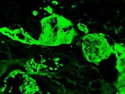
Diagnostic Biosystems FIBRINOGEN-FITC Fibrinogen-FITC
This antibody reacts with native fibrinogen and with fibrinogen fragments D, E, X, and Y. Traces of contaminating antibodies have been removed by solid phase absorption with human plasma proteins. This antibody is well suited for tissue section staining but may also be used for other applications.

Diagnostic Biosystems CD138 CD138 (EP201)
CD138, also known as Syndecan-1, is a member of the transmembrane heparan sulfate proteoglycan family, acts as an extracellular matrix receptor and is involved in many cellular functions, including cell-cell adhesion and cell-matrix adhesion. CD138 expression is found in both hematopoietic and non-hematopoietic cells. In the hematopoietic system, CD138 labels plasma cells. It is an excellent marker for plasmacytic differentiation within the spectrum of hematologic malignancy. Among non-hematolymphoid cells, CD138 reactivity is observed in many types of epithelial cells and stoma cells in both normal and tumor tissues.
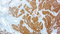
Diagnostic Biosystems CALPONIN Calponin-1
Multiple isoelectric variants of calponin have been identified but only two molecular weight isoforms exist (34kDa and 29kDa). Expression of the 29kDa form, I-calponin, is primarily restricted to muscle of the urogenital tract, whereas the higher molecular weight variant has been demonstrated in vascular and visceral smooth muscle. Calponin is a calmodulin, F-actin and tropomyosin binding protein, which is thought to be involved in the regulation of smooth muscle contraction. Calponin expression is restricted to smooth muscle cells and has been shown to be a marker of the differentiated (contractile) phenotype of developing smooth muscle.

Diagnostic Biosystems UNOVUE MOUSE Mouse/Rabbit UnoVue™ HRP/DAB Detection System
The Mouse/Rabbit UnoVue™ HRP/DAB Detection System is a non-biotin, one-step detection system suitable for demonstrating antigens in formalin-fixed paraffin-embedded tissues and cryostat sections. The UnoVue Detection System may also be used with blood smears, cytosmears, and cell preparations. The UnoVue detector kits have been developed by directly labeling anti-mouse and anti-rabbit immunoglobulins with enzymes using a proprietary tandem hyper-labelling technology. This ensures consistent and reproducible immunodetection of mouse and rabbit antibodies with a single reagent. Nuclear, cytoplasmic and membrane antigens in different types of tissues can be detected readily. The single step UnoVue Detection System enables faster staining procedures than traditional two-step methods using biotin and avidin/streptavidin conjugates, avidin/streptavidin conjugates, with significantly lower background.

Diagnostic Biosystems PEROXIDASE Universal HRP Immunostaining Kit (For Mouse and Rabbit Primary Antibodies)
This product is intended for qualitative immunohistochemistry with normal and neoplastic formalin-fixed, paraffin-embedded tissue sections, tobe viewed by light microscopy. Clinical interpretation of staining results should be accompanied by histological studies with proper controls. Patients’ clinical histories and other relevant diagnostic tests should be utilized by a qualified person (s) when evaluating and interpreting results. This kit is designed to label specific primary antibodies immunohistochemically on tissue sections. The immunohistochemical protocol defined in this brochure is just a guideline. Depending upon tissue fixation conditions, the primary antibody employed, and the user's experience, we encourage individual laboratory to optimize its own protocol. These reagents were tested and quality controlled using tissue sections, however, they can also be optimized for cell smears and cytospin preparations. Sufficient reagents are provided to run 50 to 100 tests.

Diagnostic Biosystems CYTOKERATIN CAM52 Cytokeratin (CAM5.2)
Anti-Cytokeratin (CAM 5.2) reagent has a primary reactivity with human keratin proteins that correspond to Moll’s peptides #7 and #8, Mr 48 and 52 kilodaltons (kd), respectively. Cytokeratin 7 and 8 are present on secretory epithelia of normal human tissue but not onstratified squamous epithelium. Anti-Cytokeratin (CAM 5.2) stains most epithelial-derived tissue, including liver, renal tubular epithelium, and hepatocellular and renal cell carcinomas. Anti-Cytokeratin (CAM 5.2) might not react with some squamous cell carcinomas.
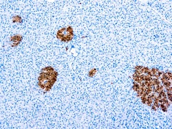
Diagnostic Biosystems NEURON SPECIFIC ENOLASE Neuron Specific Enolase
This antibody reacts with human isoenzymes of neuron specific enolase containing γ subunits. This antibody gives a specific staining in neurons of the brain, in islets of Langerhansin pancreas and in peripheral nerves of the skin. No staining with other tissue types has been seen.
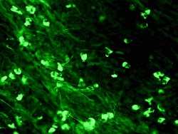
Diagnostic Biosystems IGA-FITC IgA-FITC
This antibody reacts with the alpha chain of human IgA. Traces of contaminating antibodies have been removed by solid phase absorption with human plasma proteins. This antibody is well suited for tissue section staining but may also be used for other applications.
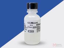
Diagnostic Biosystems CC/MOUNT CC/Mount™
CC/Mount is an aqueous mounting medium with very high refractive index. When applied to the stained tissue sections, specimens can be permanently mounted without chromogens fading. Due to the superior refractive index of CC/Mount, tissues mounted in this medium look like dehydrated specimens. No coverslipping is required with CC/Mount. However, if coverslipping is desired, dried CC/Mount can be post-mounted by using an organic based mounting medium. Advantages of CC/Mount are numerous: no coverslip, no exposure to the organic fumes, permanent storage of slides and high resolution of tissue specimens. CC/Mount is compatible with chromogens lik AEC, DAB, Fast Red, BCIP/NBT, BCIP/INT and fluorescent dyes like FITC and phycobiliproteins. The high pH of CC/Mount ensures increased stability of fluorescence.\n\nThis product is designed for permanent mounting of tissue specimens stained with peroxidase and alkaline phosphatase-based systems and various fluorescent dyes.
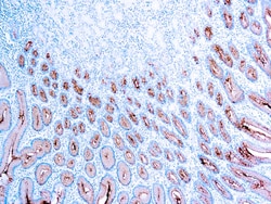
Diagnostic Biosystems MUCIN 5AC Mucin 5AC/Gastric Mucin
This antibody reacts with a 1000 kDa protein and recognizesthe peptide core of gastric mucin M1. Mucins are high molecular weight glycoproteins with 80% carbohydrates and 20% core protein. Gastric mucin M1 antigen is found in columnar mucus cells of surface gastric epithelium and in goblet cells of the fetal and precancerous colon but not in normal colon. Resurgence of gastric mucin during colonic carcinogenesis is suggestive of either reexpression of the peptide core of gastric mucin in the adult colon or changes in the glycosylation pattern of mucin, which expose the hidden M1 antigen.
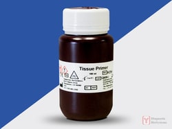
Diagnostic Biosystems TISSUE PRIMER Tissue Primer
Tissue Primer is a universal enzyme-blocking reagent to use in horse radish peroxidase (HRP) and alkaline phosphatase (AP) based immunohistochemical staining procedures done on paraffin tissue sections, frozen tissue sections and cell smears to suppress nonspecific staining due to endogenous enzyme activity.\n\nIn the evaluation of tissue specimens by IHC using HRP and AP methods, the presence of endogenous enzymes can often obscure the specific staining of the target antigen. Endogenous peroxidase and AP activity can be inhibited by the use of Tissue Primer which will suppress endogenous peroxidase, pseudoperoxidase, and alkaline phosphatase in paraffin tissue sections, frozen tissue sections and cell smears.
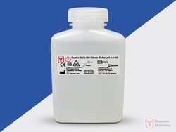
Diagnostic Biosystems QUATRO SOL I QuatroSol I 10X Citrate Buffer For Heat Induced Epitope Recovery, pH 6.0 kit/200 Runs
QuatroSol I 10X Citrate Buffer For Heat Induced Epitope Recovery, pH 6.0 kit is designed for use during the heat induced epitope retrieval (HIER) step prior to immunohistochemistry on formalin-fixed paraffin embedded tissue sections. The use of this buffer in combination with heat (often by microwave, water bath, or pressure cooker) has been shown to restore the antigenicity of proteins modified during the formalin fixation of tissue. This buffer is supplied as a 10X stock solution.\n\n